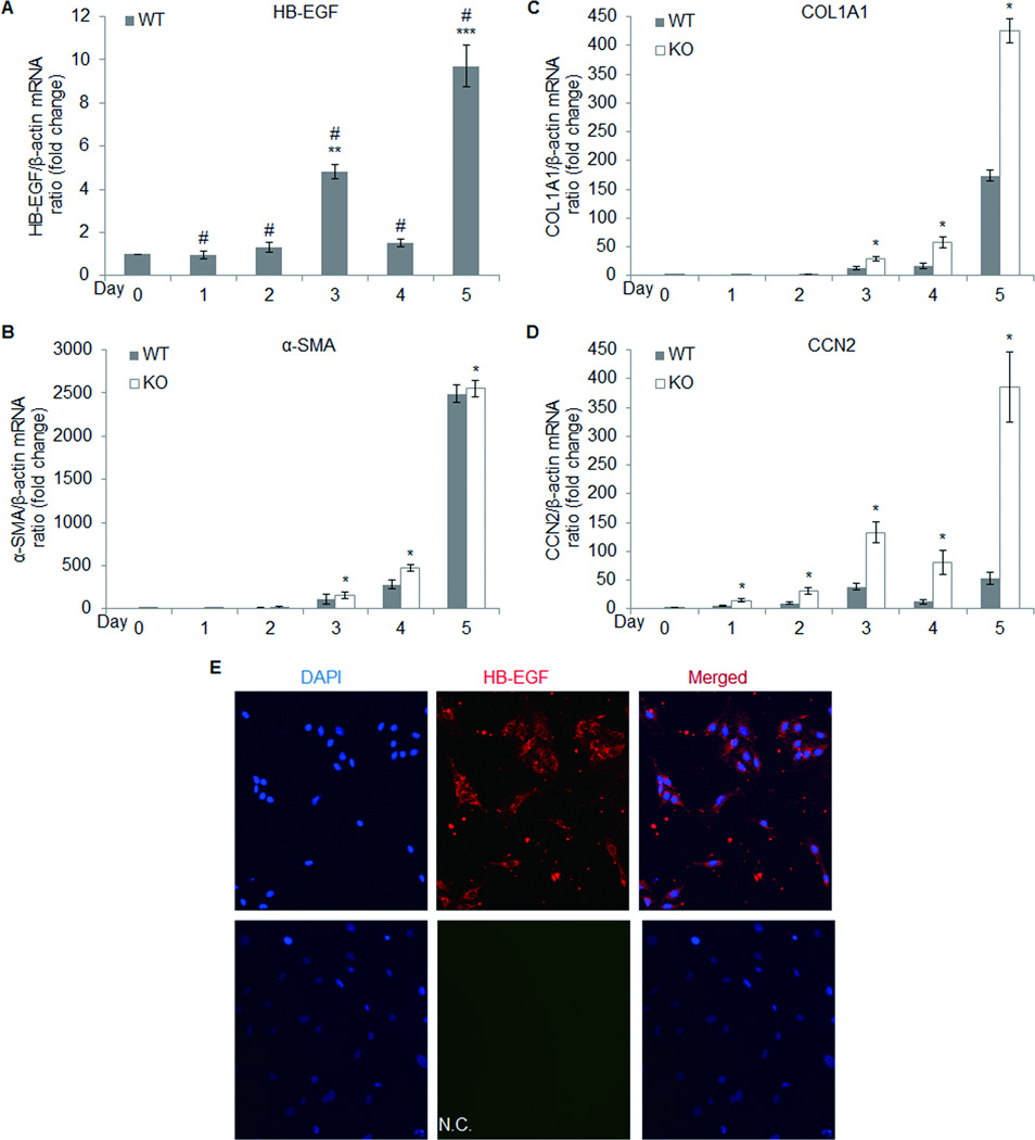
Fig. 4. Fibrogenic gene expression in primary cultured HSC.
Stellate cells isolated from untreated HB-EGF WT or KO mice were cultured for 5 days. Total cellular RNA extracted from each day of culture was subjected to quantitative real-time PCR to determine expression of HB-EGF (A), α-SMA (B), COL1A1 (C) or CCN2 (D). Wild type cells grown on coverslips for 5 days were fixed and processed to detect HB-EGF protein by immunofluorescence (E). Data are the mean ± S.D. of three experiments with triplicate determinations. # p < 0.05 vs Day 0; **p < 0.05 vs Day 1 or Day 2; ***p < 0.05 vs Day 1, 2, 3 or Day 4; *P < 0.05 vs WT. N.C., negative control or without anti-HB-EGF antibody.

