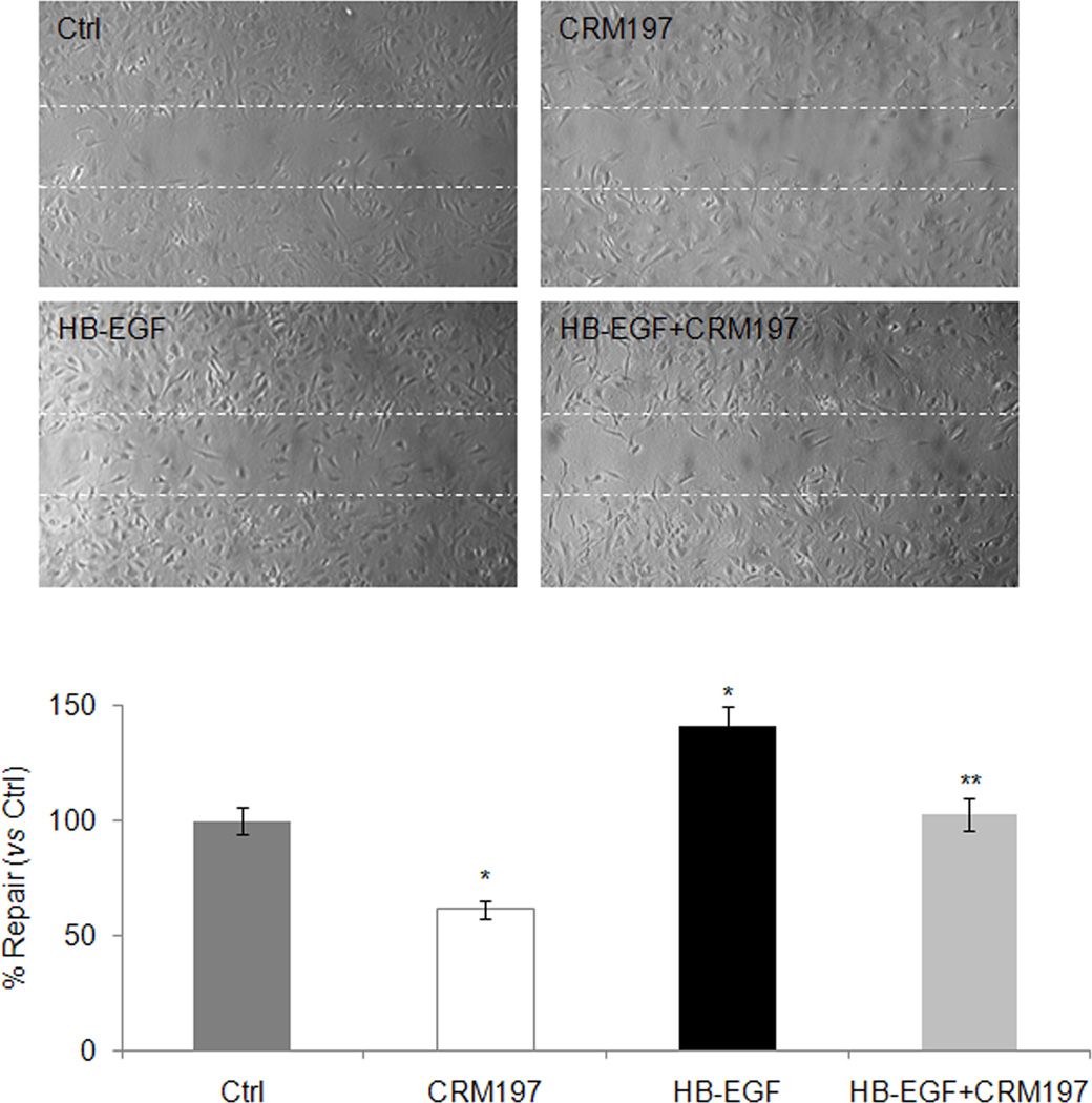
Fig. 7. Effect of exogenous recombinant HB-EGF on HSC migration.
Primary cultured HSC isolated from HB-EGF WT mice were cultured for 5 days, detached by digestion with trypsin, and plated into 6-well tissue culture plates. After pre-incubation in DMEM/F-12 containing 0.5% fetal bovine serum for 24 hours, the cultures were scratch-wounded with a 10-µl pipette tip, and incubated with 100 ng/ml recombinant HB-EGF with or without 10 µg/ml CRM197 for an additional 24 hours. The bar chart shows the number of cells in the scratched area (% versus control). Each bar represents mean ± S.D. from three experiments. Photomicrographs are representative of three experiments. Dotted line indicates wound margin. *p < 0.05 vs Ctrl; **P < 0.05 vs HB-EGF. Ctrl, control.

