Fig. 2.
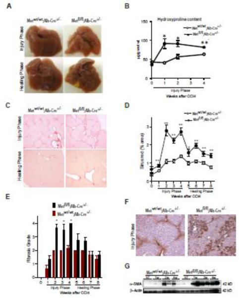
Genetic loss of c-Met receptor in hepatocytes results in excessive HSC activation and progression of CCl4-induced fibrosis. (A) Macroscopic appearance of liver. (B) Hydroxyproline content. Values are mean ± SE (n=5). (C) Representative images of Sirius red staining during injury (3 w) and healing phase (8 w) in Metfl/fl;Alb-Cre+/− and Metwt/wt;Alb-Cre+/− mice. (D) Quantification of Sirus red-positive areas using NIH ImageJ software. The data are presented as means ± SE (n=5). (E) Histological grading of fibrosis (adapted from Scheuer et al[19]). The data are presented as means ± SE (n=5). (F) Representative images of α-SMA immunohistochemistry during injury phase (3 w). (G) Western blotting of whole cell lysates prepared from Metfl/fl;Alb-Cre+/− and Metwt/wt;Alb-Cre+/− livers using anti-α-SMA. β-Actin served as a loading control. * P<0.05; **P<0.01.

