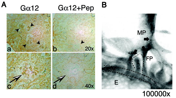
Figure 1. Endogenous Gα12 is expressed in normal mouse kidney.

(A) Immunohistochemistry of normal mouse kidney demonstrates Gα12 expression in glomeruli. Sections were probed with rabbit anti-Gα12 and visualized with Vectastain. Two magnifications (20x and 40x) are shown (panel a, b). Negative controls were performed by pre-incubating the antibody with excess blocking peptide (Gα12 + Pep). Kidney sections using the blocked Gα12 antibody showed a significant reduction in staining (panel b, d). (B) Immunoelectron microscopy shows Gα12 localizes to interdigitating foot processes (FP) and major processes (MP). Immunogold labeling and electron microscopy were performed as described in materials and methods. Magnification ~100,000x. Arrows denote gold particles. Glomerular basement membrane (GBM), foot processes (FP), fenestrated endothelium (E), and larger major processes (MP) are labeled.
