
Figure 4. Light micrographs show focal and segmental glomerulosclerosis in the juxtamedullary region of older QLα12LacZ+/Cre+ mice.
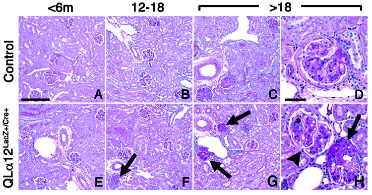
Representative light micrographs of murine juxtamedullary kidney cortex in control (top row) and QLα12LacZ+/Cre+ (bottom row) mice aged 4.5, 13, and 24 m are shown (A,E) Kidneys of mice aged <6 m, regardless of genotype, show no significant pathologic changes in glomeruli, tubulointerstitium, or vasculature. (B, F) Kidney of QLα12LacZ+/Cre+ mice (F) aged 12-18 months exhibit focal glomerulosclerosis involving juxtamedullary glomerulus (arrow). The parenchyma is otherwise well-preserved. Age-matched controls (B) show no significant pathologic changes. (C,D,G,H) Kidneys of QLα12LacZ+/Cre+ mice aged >18m show focal global (G) and segmental (H) glomerulosclerosis involving multiple juxtamedullary glomeruli (arrows), accompanied by focal interstitial mononuclear inflammation. The non-lesional glomeruli (H; arrowhead) exhibit moderate mesangial expansion by matrix and cells (*). Age-matched controls (C,D) show mild mesangial expansion but no other cortical parenchymal lesions are apparent. PAS; bar = 100 microns (left 3 columns), 50 microns (right column).