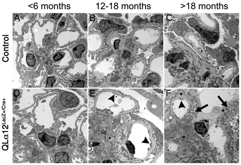
Figure 5. QLα12LacZ+/Cre+ develop foot process fusion and ultrastructural changes that worsen with age.

Transmission electron microscopy (EM) was performed on kidneys from control and QLα12LacZ+/Cre+ mice at <6m(A, D), 12-18m (B, E), and >18 m (C, F) were analyzed in a blindly and scored for severity of injury. Representative micrographs in control (top row) and QLα12LacZ+/Cre+ (bottom row) mice aged 4, 14, and 23m are shown. At <6m, both control (A) and QLα12LacZ+/Cre+ (D) show normal glomerular structure. By 12-18m, the QLα12LacZ+/Cre+ mice (E) show more signs of endothelial injury (arrowhead) and mesangial expansion (*) than controls (B). All of the oldest mice examined show significant GBM thickening but the QLα12LacZ+/Cre+ (F) show increased development of subepithelial basement membrane projections (◆) along the GBM. The podocytes have more foot process effacement and irregularity (arrow) in addition to the mesangial expansion (*) and endothelial injury arrowhead) seen the 12-18m mice compared with controls (C).
