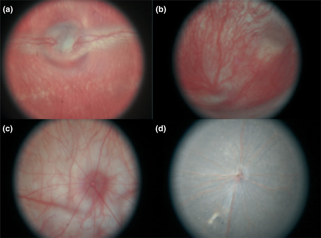
Figure 4.
(a) Image of a normal 9-month-old albino rabbit (AL = 18 mm). The optic nerve, mylinated nerve fibers, and retinal vessels are present. Deeper choroidal vessels are also present. (b) Image of a normal 3-month-old guinea pig (AL = 9 mm). (c) Image of a normal 10-week-old rat (AL = 6 mm). (d) Image of a normal pigmented 8-week-old mouse (AL = 2.5 mm).

