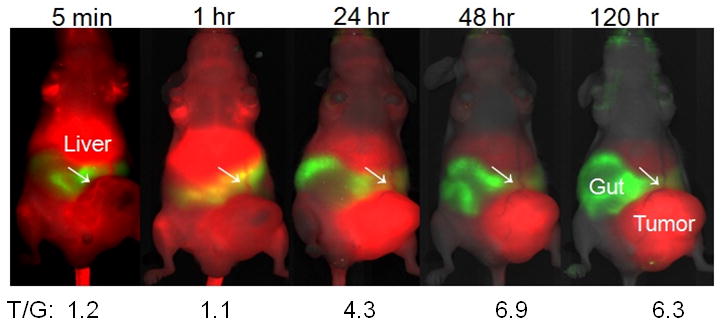
Figure 4.

Optical imaging of a mouse with human ovarian xenograft tumor. Shown are kinetic changes of Mab CC188- NIR dye IRDye 800CW conjugate in blood circulation. White arrows indicate blood vessels that supply nutrient and oxygen to the tumor. Red color indicates 800 nm channel (Mab-NIR dye probe), and green indicates 700 nm channel of gut fluorescence for contrast. The majority of the imaging probes is eliminate from circulation 24 hr post injection and completely removed from blood 48 hr after injection. T/G (Tumor /Gut) ratios are shown on the bottom of the images.
