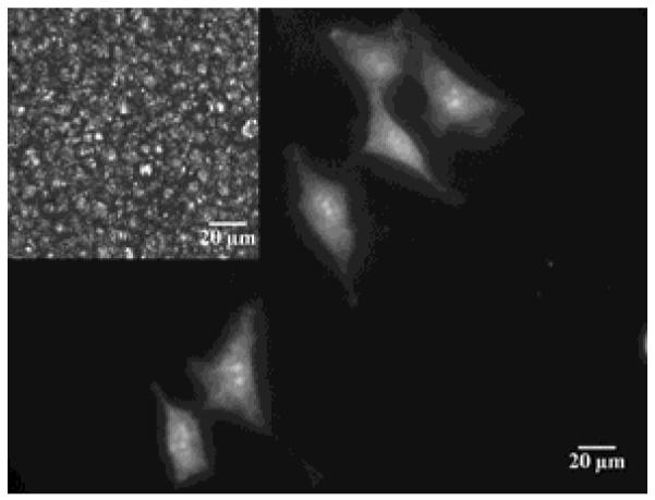
Figure 12.

Micrograph of fibroblast cells growing on a colloid-in-LC gel. The cells are stained with calcein-AM (scale bar = 20 μm) and the LC used to form the gel was E7; inset: corresponding polarized light image showing bright nematic LC domains confined by dark boundaries formed by colloids. Reproduced with permission from ref 41 (Wiley-VCH).
