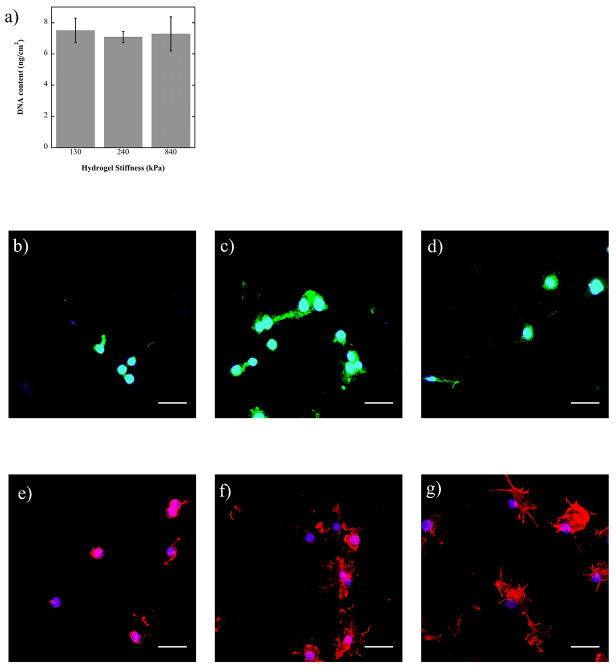
Figure 1.
a) Macrophage (RAW 264.7) attachment, measured by DNA content, after 24 hours in vitro when cultured on 130, 240 and 840 kPa PEG-RGD hydrogels (n=4). b–d) Spatial localization of αv integrins in macrophages (RAW 264.7) cultured on 130 (b), 240 (c), and 840 (e) kPa PEG-RGD gels for 48 hours. e–f) Spatial localization of F-actin in macrophages (RAW 264.7) cultured on 130 (e), 240 (f), and 840 (g) kPa PEG-RGD gels for 48 hours. Nuclei are counterstained with DAPI (blue) (b–g). Scale bar = 32 μm.

