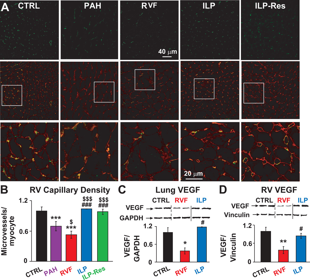
Figure 4. Preservation and even stimulation of cardiac capillary growth as well as preservation of cardiopulmonary VEGF expression by ILP.
A. Single confocal images of RV sections immunostained for CD31 (green, upper panel), overlay of CD31 (green) and wheat germagglutinin (red, middle panel) and at higher display magnification of the respective squares (lower panel). B. Quantification of RV capillary density as microvessels/cardiomyocyte. Representative immunoblots of lung (C) and RV (D) lysates labelled with anti-VEGF and GAPDH or vinculin antibodies together with Western blot analyses normalized to GAPDH or vinculin. *P<0.05 vs. CTRL; **P<0.01 vs. CTRL; ***P<0.001 vs. CTRL; $P<0.05 vs. PAH; $$$P<0.001 vs. PAH; #P<0.05 vs. RVF; ###P<0.05 vs. RVF (n=3 animals per group).

