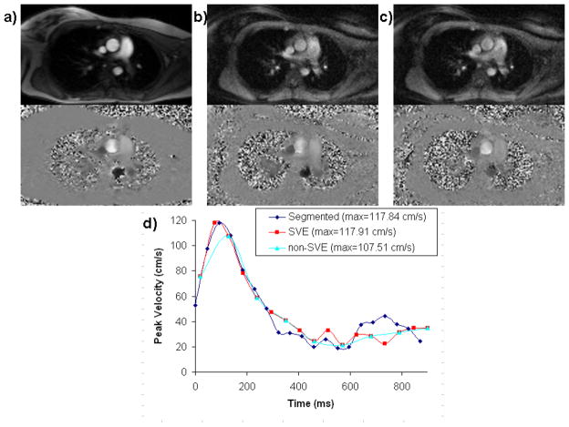
Figure 4.
Example magnitude and phase-velocity images for segmented k-space (a), real time non-SVE (b), and real time SVE (c) of the thoracic aorta in a volunteer showing typical image quality. Aortic velocity waveforms are shown from one volunteer acquired using segmented k-space, real-time SVE, and real-time non-SVE (d). Note the degradation in peak velocity when real-time acquisition is used without SVE reconstruction.

