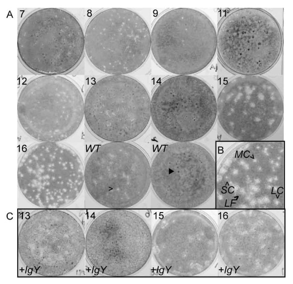
Figure 3.
Variation in plaque morphologies in IgY-selected viruses. (A) Plaque assays performed in the absence of anti-CalX polyclonal chicken IgY antiserum on IgY-resistant isolates. Passage numbers are indicated in top left corner of each image. Note the presence of multiple plaque morphologies, including small clear plaques [open arrowhead] and dark spots [filled arrowhead] in wild type [WT] as well as IgY-resistant passages. (B) Enlargement of plaque assay of passage 16 showing plaques of small clear [SC], medium clear [MC], large clear [LC] and large fuzzy [LF] morphologies. (C) Plaque assays performed in the presence of 10-3 diluted anti-CalX polyclonal chicken IgY antiserum. The variability of plaque morphologies suggests the presence of multiple variant lineages in most passages (see Additional file 1, Figure S1). Note that, except for passage 15, the constellation of different plaque types did not appreciably differ when infection took place in the presence of IgY, suggesting that no particular variant was significantly more resistant to antibody than others. Note that images were converted from color to grayscale, and brightness and contrast were adjusted to enhance plaque visibility. All image manipulations were performed in Adobe Photoshop.

