
Figure 1. Genetic deletion of β-catenin after engraftment of BCR-ABL positive disease leads to reduction of leukemia cells in blood and BM, but does not prolong survival of recipient mice.
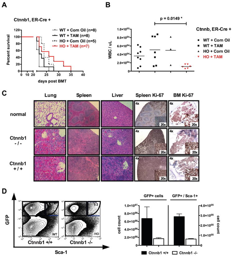
(A) After injection of 70,000 sorted GFP+ cells plus 1×10⁶ supporter cells, no significant difference in survival was evident in primary recipient mice of homozygous floxed (HO) or wildtype (WT) BM cells. β-catenin (Ctnnb1) was excised after administration of Tamoxifen (TAM) and Corn Oil served as an ‘empty control’. (B) Blood counts of moribund animals display significant differences between recipients of homozygous (Ctnnb1fl/fl Cre+) (p=.0149*) and wildtype (Ctnnb1+/+ Cre+) controls. (C) BM analysis reveals reduction in GFP+ (~3 fold reduction in Ctnnb1-/- recipients) and GFP+Sca-1+ cells (~3fold reduction in Ctnnb1-/- recipient mice). The left panel demonstrates GFP % and the right panel absolute number of GFP+ cells. (D) Organ infiltration (predominantly lung infiltration) was the cause of death in the majority of recipient mice without differential infiltration patterns between the groups investigated (displayed are representative HE-stains of each genetic group). Loss of β-catenin did not influence proliferation (Ki67) of the majority of transformed cells in spleen and BM. Error bars indicate standard deviation (SD) of 3 cohorts investigated. Excision control PCR on sorted GFP+ bone marrow cells from moribund primary recipient mice is indicated in Supplemental Figure S1.