
Figure 5. Pharmacologic inhibition of PGE2-signaling in combination with IM treatment leads to reduction of LSCs in a serial BM transplantation assay in vivo.
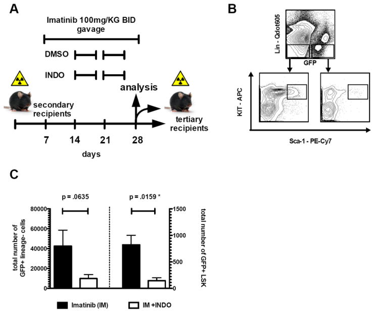
(A) Treatment schedule of secondary recipient mice (n ≥ 8 mice/cohort) injected with 150,000 GFP+Sca-1+ cells out of primary recipient mice. (B) Representative analysis plot of GFP+Lin- and GFP+LSK compartments of secondary recipient mice after treatment. (C) Co-treatment of secondary recipient mice with IM and indomethacin leads to reduction of GFP+Lin- cells (p=.0635) and to significant depletion of GFP+LSK cells (p=.0159) in the BM. GFP+Lin- cells were reduced from 42,560 (IM only group) to 9,867 (IM+INDO treated cohort). This translated into a significant reduction of GFP+LSK cells from 823 to 146, respectively. (Error bars indicate SD of GFP positive cells; n>4 per cohort investigated). Evaluation of secondary recipients after drug combination therapy is displayed in Supplemental Figure S4.