
Figure 3. CD4+ T cells are the main source of IL-10 following BBI treatment.
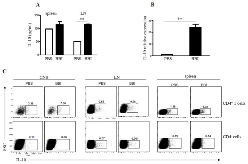
C57BL/6 mice were immunized with MOG35-55 and treated daily with BBI (1 mg/day) or PBS by oral gavage from day 0 p.i. and sacrificed on day 7 p.i. (A) LN cells were stimulated with MOG35-55 for 3 days, and concentrations of IL-10 measured in the supernatants by ELISA. (B) C57BL/6 mice (n=6 per group) were immunized with MOG35-55 and treated daily with BBI (1 mg/day) or PBS by oral gavage from the day of immunization. Mice were sacrificed on day 14 p.i.; mononuclear cells were isolated from the CNS and used to isolate mRNA. mRNA levels of IL-10 were determined by quantitative real-time PCR. Data are representative of three independent experiments. (C) Spinal cord, splenic and LN cells of immunized mice that had been treated for 14 days p.i. with BBI were isolated and analyzed for intracellular production of IL-10 by flow cytometry. ** p < 0.01. Data are representative of three independent experiments.