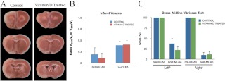
Fig. 7.
Effect of vitamin D3 injections on infarct volume. Animals were subject to MCAo and euthanized 5 d later. A, Representative slices of TTC-stained sections depict the rostrocaudal extent of the infarct in control and vitamin D3-treated animals. B, Histograms depict infarct volume normalized to the contralateral side. Infarct volume was not affected by acute vitamin D3 treatment in the cortex or the striatum. Bars represent mean ± sem. C, Performance on sensory motor task before and after ischemia. Animals were tested before and after ischemia on the vibrissae-evoked forelimb placement task. Histogram depicts mean (±sem) correct responses on the cross-midline vibrissae-elicited forelimb placement task for each forelimb. Postischemic performance was significantly affected on the left and right limb. However, vitamin D3 treatment had no affect on the performance of either limb. Data were analyzed by two-way ANOVA for pre-/postischemia and vitamin D3 treatment; b, Main effect of ischemia. Group differences were considered significant at P ≤ 0.05; n = 4–5 in each group.Vctx, Volume cortex; Vstr, volume striatum; Vc, volume contralateral hemisphere.

