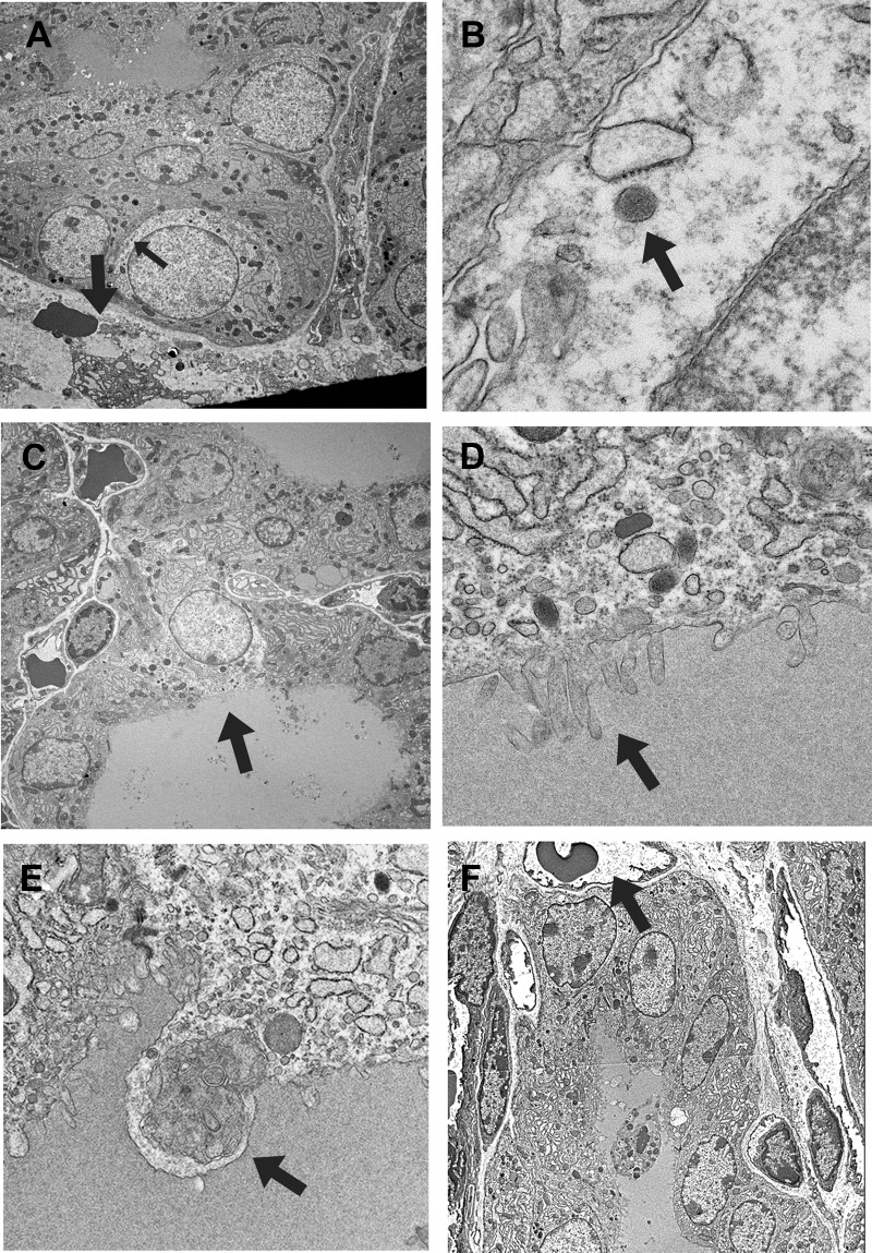
Fig. 2.
EM analysis of the proliferative area of the thyroid. A, Clear cell that retains characteristics of C cells such as being surrounded by other cells and the presence of neuroendocrine granules (representative, indicated by a small arrow). The thick arrow indicates a capillary vessel with erythrocytes. B, High magnification of a neuroendocrine granule shown in A (indicated by an arrow). C, Clear cell that retains follicular cell characteristics such as those facing the lumen and the presence of microvilli at the apical side of the cell (indicated by an arrow). D, High magnification of microvilli shown in C (indicated by an arrow). E, Cell undergoing autophagy. Various cytosolic vesicles are found inside lysosome (shown by an arrow). F, Dying cells and cells surrounding them, which are about to form a lumen and a follicle, respectively. An arrow indicates capillary vessel with erythrocytes. Original magnification: ×1000 (A, C, and F), ×20,000 (B), ×10,000 (D), and ×5000 (E).

