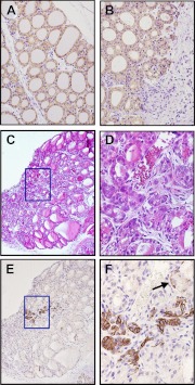
Fig. 6.
Cytokeratin expression after PTx. A and B, Immunohistochemistry for Krt19 of intact (A) and 7 d after partially thyroidectomized thyroids (B). C and D, H&E staining. E and F, Immunohistochemistry for Krt14 counterstained with hematoxylin. C and E are from serially prepared sections. Box in C corresponds to the area where Krt14 is expressed in E, and the magnified images of the boxed areas are shown in D and F, respectively. The arrow in F indicates that only basal cell cytoplasm is positively stained for Krt14 (basal cell pattern), whereas the upper cells (luminar cells) are negative for Krt14 expression. Original magnification: ×100 (C and E), ×200 (A and B), and ×400 (D and F).

