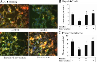
Fig. 6.
Prolonged exposure of hepatocytes to insulin decreases mitochondrial membrane potential in a cholesterol synthesis-dependent manner. Hepa1c1c7cells (A and B) or primary mouse hepatocytes (C) were treated with either the vehicle solution or insulin (5 nm) in the presence of simvastatin (10 nm) for 16 h as indicated (n = 3). Mitochondrial membrane potential was then either visualized by using fluorescent microscopy (A) or quantification of fluorescence density (B and C) as detailed in Materials and Methods. Results represent mean ± se of three independent experiments. **, P < 0.01 vs. no insulin control. #, P < 0.05 and ##, P < 0.01 vs. insulin alone.

