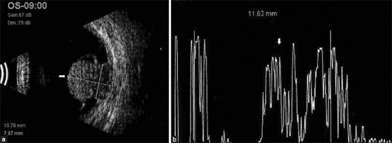
Figure 1.

(a) B-scan ultrasound showing a large mushroom-shaped mass lesion (arrow) noted nasally with sub-retinal fluid noted surrounding the mass lesion. (b) A-scan ultrasound of the same lesion showing internally medium reflectivity (arrow) in the lesion area
