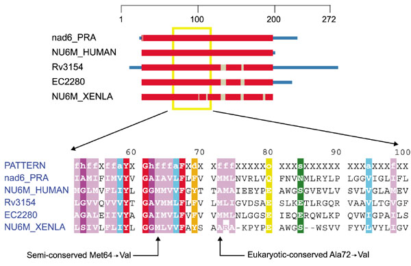
Figure 3.

Domain diagram and partial alignment for NADH dehydrogenase, subunit 6 (ND6). The columns indicated with arrows at the bottom are positions 64 and 72 of NU6M_HUMAN. Sequences are from M. tuberculosis (Rv3154), Escherichia coli (EC2280), X. laevis mitochondrion (NUAM6_XENLA), H. sapiens mitochondrion (NU6M_HUMAN) and R. americana mitochondrion (nad6_PRA). Colors are as in Figure 1.
