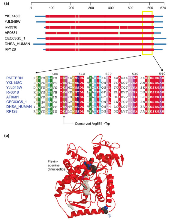
Figure 7.

Structures of the flavoprotein subunits of succinate dehydrogenase and fumarate reductase. (a) Domain diagram and alignment for the flavoprotein subunit of succinate dehydrogenase (SDHA). The column indicated by the arrow on the bottom corresponds to position 554 of DHSA_HUMAN. Sequences come from S. cerevisiae (YKL148W and YJL045W), M. tuberculosis (Rv3318), A. fulgidus (AF0681), Caenorhabditis elegans (CEC03G5_1), H. sapiens (DHSA_HUMAN) and R. prowazekii (RP128). Colors are as in Figure 1. (b) Ribbon diagram of the flavoprotein subunit of fumarate reductase (E. coli) - a very close homolog of SDH Fp (Z-score 175.8). The structure comes from the Protein Data Bank (code: 1FUMA). Flavin adenine dinucleotide can be seen (in space-filling representation) in the middle of the structure and Thr494 is similarly indicated at the right-hand end of the bottom-most helix (1). Arg554 occupies this position in DHSA_HUMAN.
