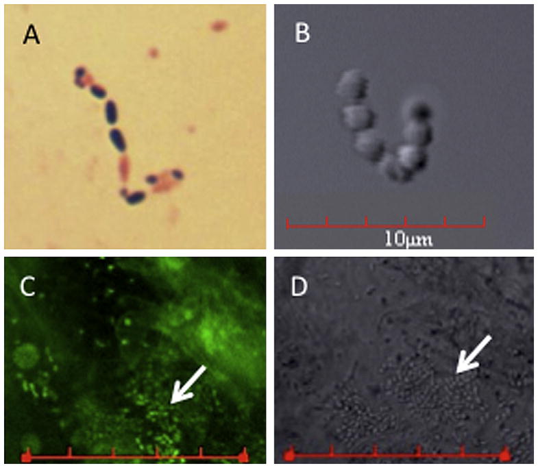
Figure 2.

Microscopic analysis of Enterococcus isolates. A: Gram stain of the E633 E faecalis isolate (IOL isolate from this case). B: Differential interference contrast photomicrograph taken of IOL showing coccus-shaped bacteria. C: Confocal laser scanning microscopic image of the removed IOL that had been incubated with a DNA-fluorescent dye. D: The differential interference contrast image of the same region of the IOL shown in C. These data support that strain E633 formed a biofilm on the IOL (bars in C and D = 30 μm; arrows indicate bacterial biofilm).
