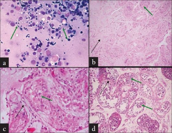
Figure 1.

(a) FNAC from testis showing normal spermatogenesis. (b) Testis biopsy showing patchy areas of atrophy and hyalinization of seminiferous tubules (dashed arrow), interspersed tubules showing normal spermatogenesis including mature spermatozoa (green arrow). (c) Testis biopsy showing hyalinized and thickened seminiferous tubules with basement membrane thickening (black dashed arrow); some tubules showed the presence of spermatogenesis including mature spermatozoa (green arrow). (d) Testis biopsy showing mild basement membrane thickening and peritubular fi brosis (black dashed arrow) and maturation arrest till spermatid stage (green arrow)
