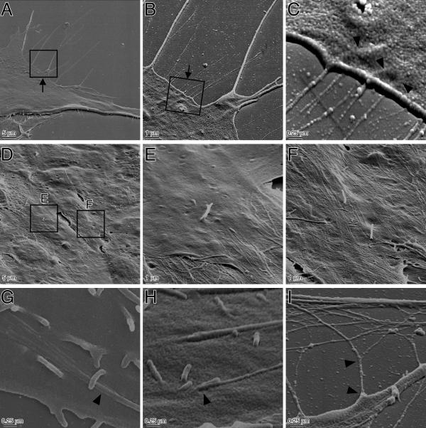
Figure 1.
Scanning electron micrographs of NIH3T3 fibroblasts with a tilt angle 60°. (A-C) Exponentially growing NIH3T3 cells were seeded onto serum-coated glass coverslips at relatively low density (1 × 10³ cells/cm2) thereby avoiding cells to contact each other. After 48 hours, cells were fixed with 2% glutaraldehyde in PBS(+), and prepared for SEM as described in Materials and Methods. (A) SEM image of NIH3T3 cell at low magnification. Note several micropodia, but not ultramicroextensions, at this magnification. (B) Higher magnification view of area indicated by square in A. Note that ultramicroextensions can be detected at this magnification. (C) Higher magnification view of area indicated by square in B. Sample was observed from the direction indicated by arrow in B. Note the diameter of ultramicroextensions is less than 0.05 μm and some of the ultramicroextensions appear to originate from a ridge of the cell body (arrowhead). (D-F) Cells were seeded onto serum-coated glass coverslips at 1 × 10⁴ cells/cm2. After 1 week, cells were fixed with 2% glutaraldehyde in PBS(+) and prepared for SEM. (D) SEM image of NIH3T3 cell at low magnification. Note that cells are attached to each other and about half of the cell surface is not smooth. (E and F) Higher magnification view of area indicated by square in D. Note the numerous and parallel rather thin and long microextensions covering some of cell surface. (G-I) High magnification view of an NIH3T3 cell showing the origins of ultramicroextensions. They appear to grow from the tip of microextension in a broom-like fashion (G), from the cell surface like microvilli (H), or branch off from microextensions or ultramicroextensions (I).

