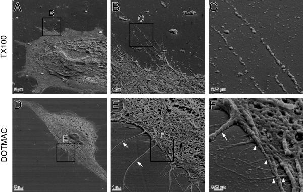
Figure 2.
(A) Scanning electron micrographs of NIH3T3 fibroblast extracted with Triton X-100 or DOTMAC at a tilt angle 60°. (A-C) NIH3T3 cells on glass coverslips were rinsed with PBS(+) and fixed with 4% paraformaldehyde in PBS for 10 min at room temperature. Cells were then extracted with 0.2% Triton X-100 for 5 min at room temperature, and prepared for SEM. (B) Higher magnification view of area indicated by square in A. Note that some of microextensions appeared to be lost. (C) Higher magnification view of area indicated by square in B. Note the fragmentation of microextensions. (D-F) Cells were fixed with the DOTMAC/PFA method and prepared for SEM. (E) Higher magnification view of area indicated by square in D. Note that lipid plasma membrane appeared to be lost and micropodia (white arrow) and microvilli (black arrow) are well preserved. (C) Higher magnification view of area indicated by square in E. Note that origins of ultramicroextensions (arrowhead) were exposed. Some of ultramicroextensions appear to grow out from the inner space of cytoplasm.

