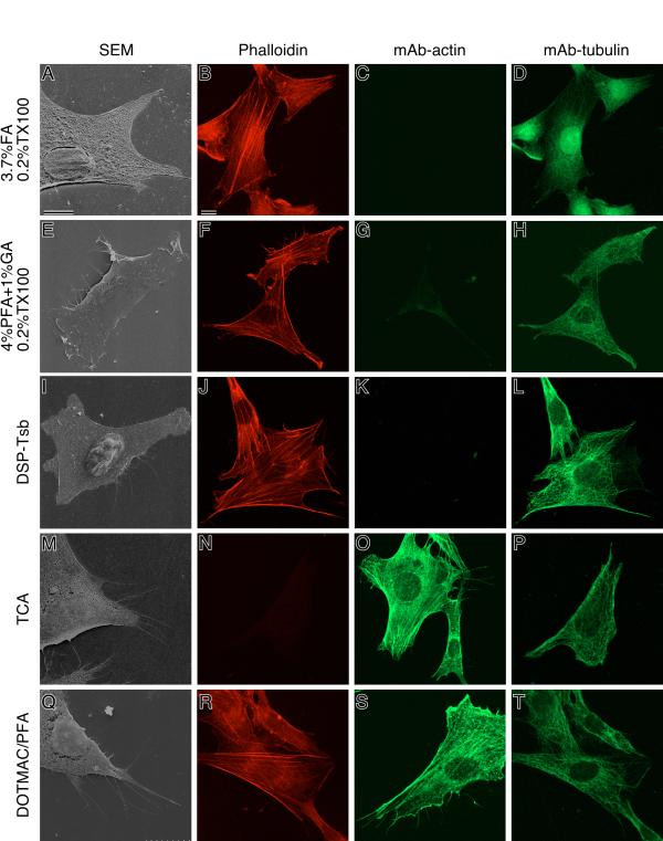
Figure 3.
Scanning electron and confocal laser scanning micrographs of NIH3T3 cells fixed with various procedures. (A, E, I, M, and Q) SEM image of NIH3T3 cells shown with no tilt angle. Cells were double labeled with TRITC-phalloidin (B, F, J, N, and R) and anti-β-tubulin monoclonal antibodies (T4026), which was detected using a secondary antibody conjugated to FITC (D, H, L, P, and T). Note that the fluorescence intensity of each samples varied as summarized in Table 2. Images are shown at optimum fluorescence intensity. Cells were also stained with anti-β-actin monoclonal antibodies (A5441), followed by FITC-conjugated anti-mouse IgG (C, G, K, O, and S). Cells were rinsed with PBS(+) and fixed with 3.7% FA/0.2% TX100 (see Table 1, A-D), 4% PFA+1% GA/0.2% TX100 (see Table 1, E-H), DSP-Tsb (see Table 1, I-L), TCA (see Table 1, M-P), or DOTMAC/PFA (see Table 1, Q-T). Bars, 10 μm.

