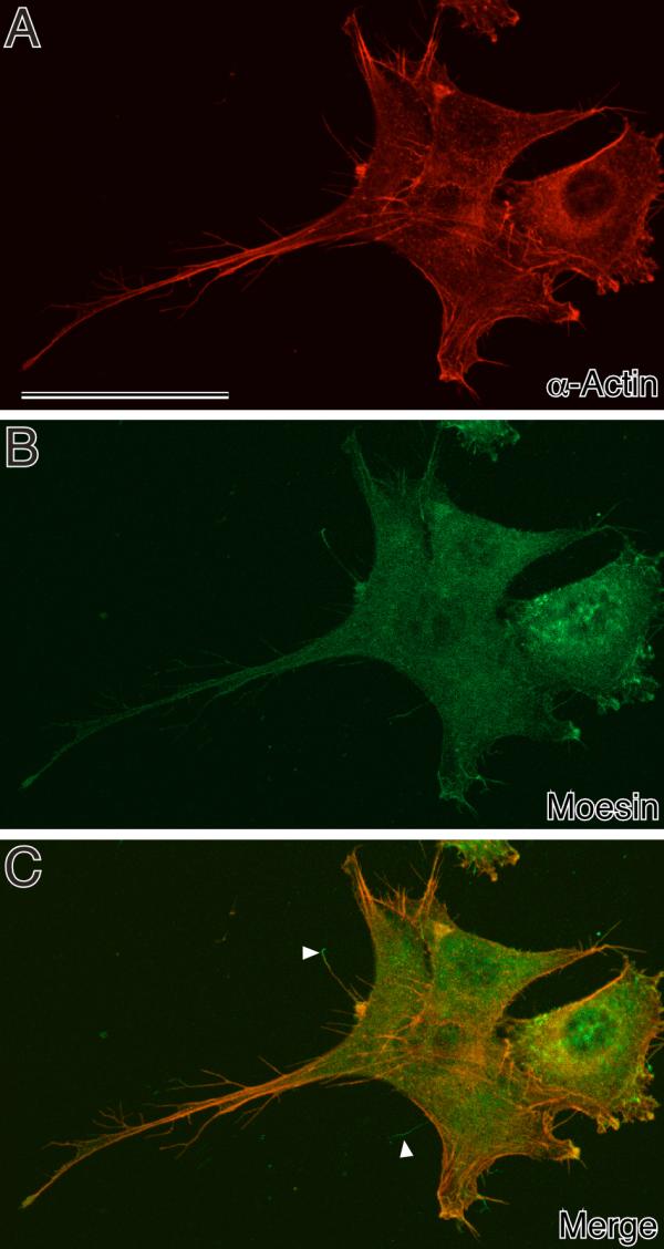
Figure 5.

Double staining of actin and moesin in NIH3T3 cells fixed with DOTMAC/PFA shown at high magnification. Note that branched micropodia were well preserved by the DOTMAC/PFA fixative and stained with both β-actin monoclonal and moesin polyclonal antibodies. Some of the moesin-positive micropodia were β-actin-negative (arrowhead). Bars, 50 μm.
