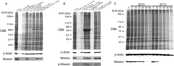
Figure 6.
SDS-polyacrylamide gel electrophoresis of extracted polypeptides from NIH3T3 cells after fixation and/or permeabilization. Subconfluent cultures of cells were rinsed with PBS(+), fixed and/or extracted as indicated. After rinsing with PBS, the material remaining on the culture dish was extracted with SDS sample buffer. The dissolved and undissolved material was collected and heated at 95°C for 10 min and resolved by SDS-PAGE on a 9% polyacrylamide gel. Polypeptides were stained with Coomassie Brilliant blue (CBB). β-Actin, moesin, and threonine558-phospho-moesin were detected by immunoblot analysis. (A) Cells were extracted with the extraction buffer as indicated in the figure for 5 min at room temperature. In the last lane to the right, cells were fixed with 1% PFA in PBS for 20 min at 4°C after extraction. (B) Cells were fixed and/or extracted with the fixative indicated in the figure as shown in Table 1 except that 3.7% formalin was used instead of 3.7% formaldehyde in lane 7. (C) Cells were fixed with 4% PFA in PBS(+) for the indicated time period (min) at 25°C or 37°C, rinsed with PBS, and then extracted with SDS sample buffer. Note that moesin is more readily irreversibly crosslinked than actin. Other polypeptides appeared to be same as moesin.

