Fig. 6.
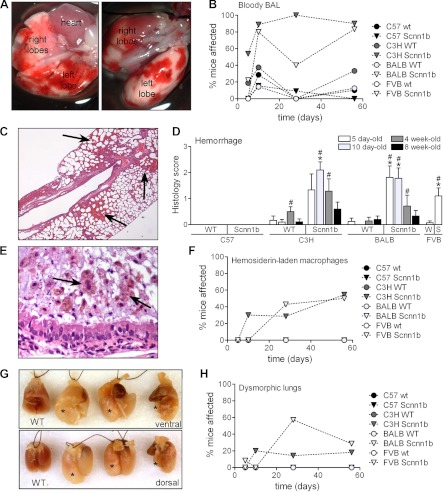
Pulmonary hemorrhage, pulmonary hemosiderosis, and dysmorphic lungs in congenic Scnn1b-Tg mouse strains. A: representative images of patchy hemorrhage observed at dissection in 10-day-old C3H/HeN Scnn1b-Tg mice, ventral and dorsal side, respectively. B: incidence of bloody BAL in congenic WT and Scnn1b-Tg strains (n = same as Fig. 4). C: representative photomicrograph of lung histology [hematoxylin and eosin (H&E) stain] in 10-day-old C3H/HeN Scnn1b-Tg mice. Arrows, areas of parenchymal hemorrhage. D: semiquantitative histopathology score for pulmonary hemorrhage. n, Same as Fig. 3. *P < 0.05 vs. WT littermates, #P < 0.05 vs. C57BL/6N of the same genotype. E: representative photomicrograph of infiltrating inflammatory cells encased in a mucus plug in the airways of a 10-day-old C3H/HeN Scnn1b-Tg mouse H&E stain. Hemosiderin-laden macrophages exhibiting typical brown granular inclusions are present (arrows). F: incidence of hemosiderosis in Scnn1b-Tg mice and WT littermates at different time points. n, Same as Fig. 3. G: representative image of dysmorphic lungs in 3 adult BALB/cJ Scnn1b-Tg mice compared with WT age-matched control (WT). *Overdistended (left and center) or atelectatic (right) lobes. H: incidence of dysmorphic lungs in Scnn1b-Tg mice and WT littermates at different time points. n, Same as Fig. 3.

