Figure 4.
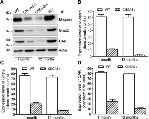
CNGA3−/− mice show reduced expression of cone-specific proteins. Western blot detection was performed using retinal membrane protein extracts prepared from CNGA3−/− and WT mice at 1 and 12 months to determine the expression of M-opsin, Gnat2, and CAR. Actin was included as a loading control. (A) Shown are the representative images of Western blot detection. Densitometric analysis of Western blot detection of M-opsin (B), Gnat2 (C), and CAR (D). Data are presented as mean ± SEM of measurements from four to five independently performed experiments using retinas from four to six mice (*P < 0.05).

