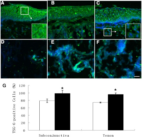
Figure 1.
Immunofluorescence staining of TSG-6 in normal and CCh conjunctiva and Tenon's capsule. One representative normal (A, D) and two representative CCh (B, C, E, F), conjunctival tissue (A, C), and Tenon's capsule (D–F) were subjected to immunofluorescence staining using a TSG-6–specific antibody. More positive immunostaining to TSG-6 was noted in CCh than in normal conjunctival stroma (B, C) and Tenon's capsule (E, F). Nuclear counterstaining was performed by Hoechst 33342. All images except those in the insets in (A) and (C) were taken at the same magnification. Scale bar, 50 μm. The percentage of TSG-6–positive cells in the subconjunctival tissue and the Tenon's capsule of normal (□) and CCh (■) samples (n = 4) were quantified and compared (G). *P < 0.01 versus normal.

