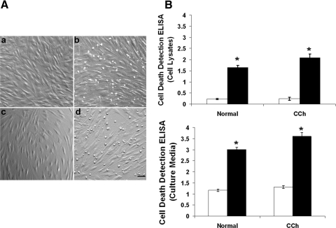
Figure 7.
Apoptosis promoted by TSG-6 knockdown. Both normal and CCh fibroblasts were cultured in DMEM with 0.5% FBS for 48 hours and were transfected with TSG-6 siRNA for another 48 hours while IL-1β was added for the last 24 hours. (A) Compared with the control treated with scRNA (a, normal fibroblasts; c, CCh fibroblasts), TSG-6 siRNA caused more detached round cells in normal (b) and CCh fibroblasts (d). All images were taken at the same magnification. Scale bar, 100 μm. Compared with scRNA (□), the extent of cell apoptosis was also significantly increased by TSG-6 siRNA (■) (*P < 0.01) in cell lysates (B, top) and in culture media (B, bottom) in normal and CCh fibroblasts.

