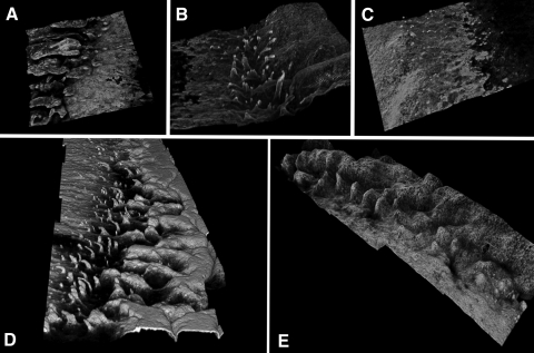
Figure 3.
(A–C) 3D confocal reconstructions of different areas of the limbus from the same subject (see Supplementary Movies S1–S3, http://www.iovs.org/lookup/suppl/doi:10.1167/iovs.11-8524/-/DCSupplemental). (D) 3D reconstruction of a limbal region showing an extensive finger-like pattern. (E) 3D reconstruction of a limbal region showing an undulating and irregular palisade pattern.

