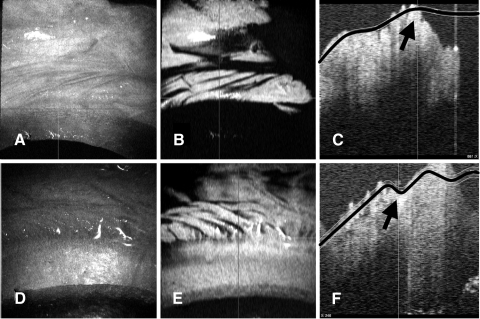
Figure 9.
Example of possible areas of clipped palisades. (A, D) The en face view shows the cut edge that is not perpendicular to the surface of the cornea. From this angle it does not appear that there has been any clipping. (B, E) C-mode reconstructions at the level of the palisades showing palisade structures coming right up to, and ending abruptly at, the cut edge. (C, F) Vertical orthogonal views of the same section showing the cut edge of the cornea. The planes of the C-mode display are marked in black, and the area of the cut edge is highlighted with a black arrow. These cuts are stepped in two stages.

