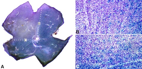
Figure 2.
Regional loss of βGEO activity occurs in 10-month-old Bax−/− DBA/2JR3/R3 mice. Cells with βGEO activity appear blue, whereas Nissl-stain appears violet. (A) Whole-mounted retina, showing a wedge-shaped region devoid of βGEO activity (defined by white hashed lines). Regions (B) and (C) in borders of the wedge are shown in higher magnification. Although cells have stopped expressing βGEO, they are still present in the wedge region. Scale bar for (B) and (C): 50 μm.

