Figure 4.
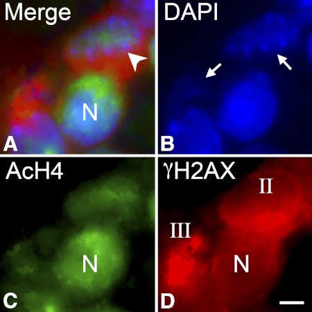
Apoptotic cells exhibit reduced staining for acetylated histone H4. Immunofluorescent double labeling of cells in the ganglion cell layer of a DBA/2J mouse. Frozen sections were double labeled with antibodies against the histone variant γH2AX to identify dying cells, acetylated histone H4 (AcH4), and counterstained for DNA using DAPI. (A) Merged image of all three labels in a section. (B–D) Individual channels for DAPI, AcH4, and γH2AX, respectively. Representative stages I, II, or III nuclei (N), based on the γH2AX labeling, are indicated. Stage I cells exhibit strong labeling for AcH4, whereas the single stage III cell in the image has virtually no AcH4 present. Stage II cells show some AcH4 staining, but it is typically intermediate between stage I and stage III labeling. Scale bar: 5 μm.

