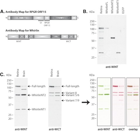
Figure 2.
(A) Antibody maps for Rpgr and whirlin, and analysis of whirlin expression in the mouse retina. Top: illustration of the RpgrORF15 isoform structure and the location of domains used to generate polyclonal antibodies. S1 antibody detects all Rpgr isoforms; ORF15 detects only RpgrORF15. Bottom: schematic representation of the whirlin long isoform structure and location of the domains used to generate polyclonal antibodies against the N-terminal and C-terminal ends. WiNT detects the long whirlin isoform and any potential short, N-terminal isoforms; WiCT also detects the long whirlin isoform and the previously reported short, C-terminal isoforms. (B, C) Characterization of whirlin antibodies and analysis of whirlin expression in the mouse retina and brain. (B) Immunoblot analysis of retinal homogenate and cell lysate from HEK293 cells transfected with the designated whirlin isoform. (C) Immunoblot analysis of retinal and cerebral homogenate from C57BL/6 wild-type mice. The WiNT antibody detects three major bands of approximately 110 kDa, 60 kDa, and 34 kDa in both the retina and the brain. We also detected an additional ∼85-kDa band in the brain that is not consistently detected in the retina and has not been identified. The WiCT antibody detects three major bands of approximately 110 kDa, 70 kDa, and 60 kDa in both the retina and the brain, with an additional ∼40-kDa band detected only in the brain. See Table 1 for GenBank accession numbers and a description of each known whirlin variant. Artificial colorization and merging of the images confirms that only the 110-kDa, long whirlin isoform is detectable by both antibodies and that all short N-terminal and C-terminal isoforms are detected only by their respective antibodies.

