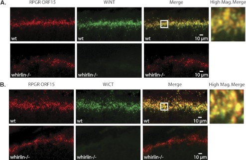
Figure 4.
Colocalization of whirlin and RpgrORF15 in the mouse retina by immunohistochemical analysis. (A, top) Double staining of C57BL/6 wild-type retina with Rpgr ORF15 polyclonal antibody (red) and WiNT polyclonal antibody (green). Bottom: double staining of whirlin knockout retina with Rpgr ORF15 polyclonal antibody (red) and WiNT polyclonal antibody (green). (B, top) Double staining of C57BL/6 wild-type retina with Rpgr ORF15 polyclonal antibody (red) and WiCT polyclonal antibody (green). Bottom: double staining of whirlin knockout retina with Rpgr ORF15 polyclonal antibody (red) and WiCT polyclonal antibody (green). (A, B) The boxed region on the merged images indicates the region shown at right at higher magnification. The higher magnification merged images indicate that both whirlin antibodies partially colocalize with RpgrORF15 in the vicinity of the photoreceptor-connecting cilia.

