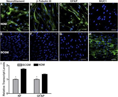
Figure 4.
Induction of neural cell differentiation from TMSCs. Immunofluorescent staining on neural markers neurofilament, β-tubulin III, GFAP, and TMSC marker MUC1 shows different expression between TMSCs in NDM (A–D) for neural induction and in SCGM (E–H) to stem cell maintenance. DAPI stains nuclei blue. Bars: 50 μm. (I) Neural markers NF and GFAP from TMSCs in SCGM and in NDM for neural induction were quantified by qRT-PCR. Error bars show SD of triplicate analyses. *P < 0.05 (n = 3, Student's t-test). GFAP, glial fibrillary acidic protein; NF, neurofilament.

