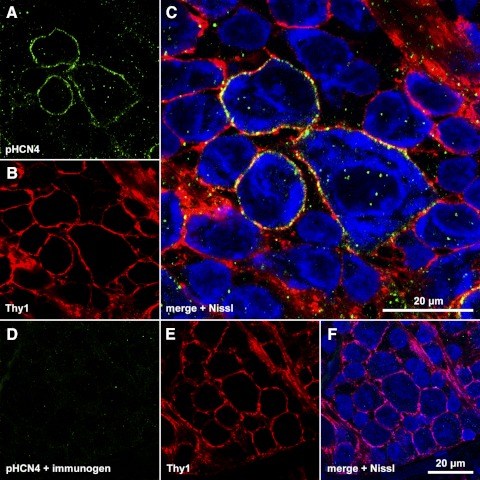
Figure 3.
Colocalization of Thy1 and HCN4 in ganglion cell layer somata. (A–C) Single optical section through the ganglion cell layer of a retina incubated in polyclonal anti-HCN4 primary and DyLight 488-conjugated secondary, anti-Thy1 primary, and DyLight 549-conjugated secondary, and Nissl stain. Fluorescence from DyLight 488 (A), DyLight 549 (B), and Nissl (C) assigned to the green, red, and blue color channels, respectively, and merged in (C). (D–F) Single optical section through the ganglion cell layer of a different portion of the same retina, processed as in (A–C) except that the anti-HCN4 primary was preincubated with immunogen. The scale bar in (C) (20 μm) applies to (C) alone; the scale bar in (F) (20 μm) applies to (A, B, D–F).

