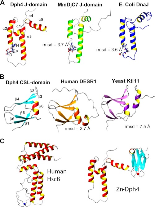
FIGURE 5.
Comparison of Dph4 subdomains with representative structures of similar folds. A, J-domain of Dph4, MmDjC7 (PDB code 1WJZ), and E. coli DnaJ (PDB code 1XBL) (top). B, CSL-domain of Dph4, human DESR1 (PDB code 2JR7) and yeast Kti11 (PDB code 1YOP) (middle). The r.m.s. deviations among the domains are indicated. The signature HPD motif of J-domains is represented in a ball and stick model. C, comparison of full-length human Dph4 and human HscB.

