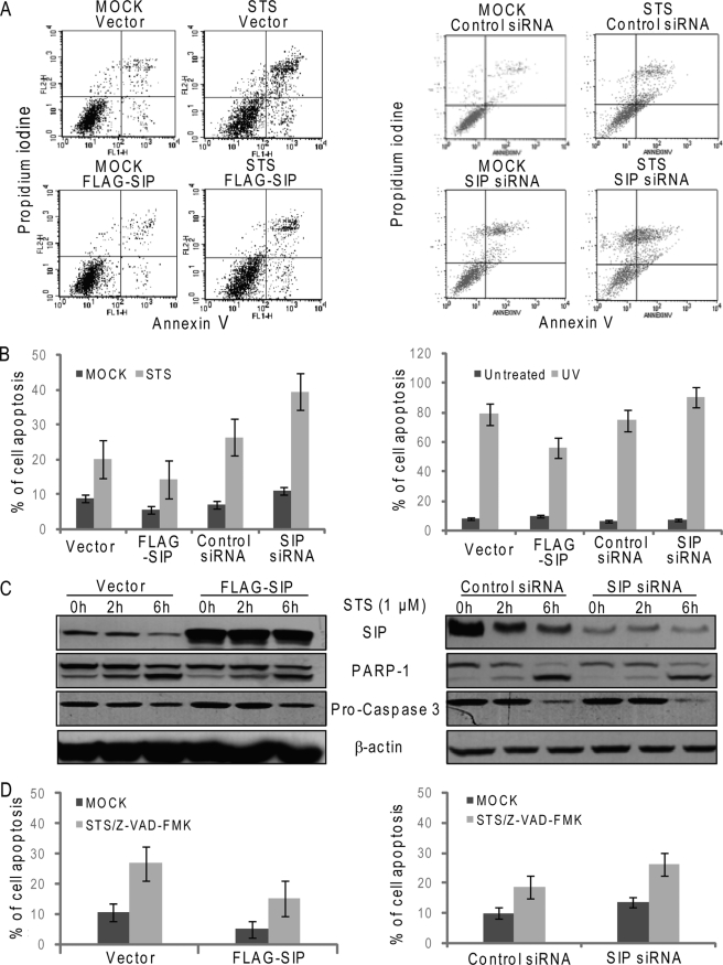
FIGURE 1.
SIP inhibits apoptosis through caspase-independent mechanism. A, FACS analysis of apoptosis by double staining with annexin V and propidium iodide. U2OS cells were transfected with SIP expression construct or SIP siRNA. Forty-eight hours after transfection, the cells were untreated (control) or treated with STS (6 h), labeled with annexin V-FITC and PI, and analyzed by flow cytometry. Representative cytofluorometric plots are shown. B, FACS analysis of apoptosis in MCF-7 cells (left panel) and U2OS cells treated with UV irradiation (right panel). MCF-7 cells described in A or U2OS cells unexposed (control) or exposed to 40 J/m2 UV for 1 min, 7 h later, were labeled with annexin V-FITC and PI and analyzed by flow cytometry. The values are expressed as percentages of annexin V-positive versus total cells. The data are the means ± S.D. from triplicate experiments. C, Western analysis of apoptosis. Total cell lysates from U2OS described in A were prepared, and the expression levels of SIP and activated PARP-1 and caspase-3 were examined by immunoblotting. β-actin was used as a loading control. D, FACS analysis of apoptosis. U2OS described in A in the absence or presence of Z-VAD-FMK were untreated (control) or treated with STS (6 h), labeled with annexin V-FITC and PI, and analyzed by flow cytometry. The values are expressed as percentages of annexin V-positive versus total cells. The data are the means ± S.D. from triplicate experiments.

