FIGURE 3.
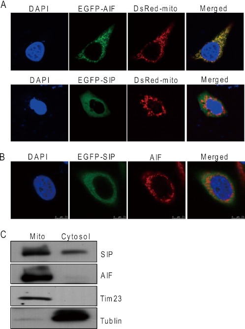
Subcellular localizations of SIP and AIF. A, subcellular localizations of SIP protein. pEGFP-AIF and DsRed-mito or pEGFP-SIP and DsRed-mito were cotransfected into HeLa cells. EGFP and DsRed fluorescence were visualized by fluorescence microscopy. 4,6-Diamidino-2-phenylindole dihydrochloride (DAPI) staining was also included to visualize the cell nucleus. B, subcellular colocalization of SIP and AIF. HeLa cells were transfected with pEGFP-SIP. Twenty-four hours after transfections, EGFP fluorescence and rhodamine staining of AIF were visualized by fluorescence microscopy. C, analysis of subcellular localization of SIP by subcellular fractionation. HeLa cells were subjected to subcellular fractionation, and immunoblotting was performed with cytoplasm (Cytosol) and mitochondrial (Mito) fractions. Tublin and Tim23 were used as cytosolic and mitochondrial marker proteins, respectively.

