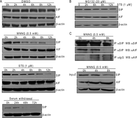
FIGURE 6.
SIP is degraded in response to apoptosis stimulus. A, Western blotting analysis of SIP expression during DMSO, STS, MNNG, or serum withdrawal-induced apoptosis. HeLa cells treated with DMSO, STS, MNNG, or serum withdrawal were collected at the indicated times. Cell lysates were prepared and analyzed by Western blotting for SIP, AIF, or β-actin. B, the degradation of SIP triggered by apoptosis stimulus can be blocked by MG132. HeLa cells were first treated with MG132 for 1 h and then treated with STS at the indicated times. Cell lysates were prepared and analyzed by Western blotting for SIP, AIF, or β-actin. C, interaction between SIP and AIF during apoptosis. U2OS cells were treated with MNNG at the indicated times. Whole cell lysates were first immunoprecipitated (IP) with anti-SIP or anti-IgG and then immunoblotted with anti-AIF or anti-SIP. Inputs from each sample were immunoblotted with anti-SIP, anti-AIF, or anti-β-actin.

