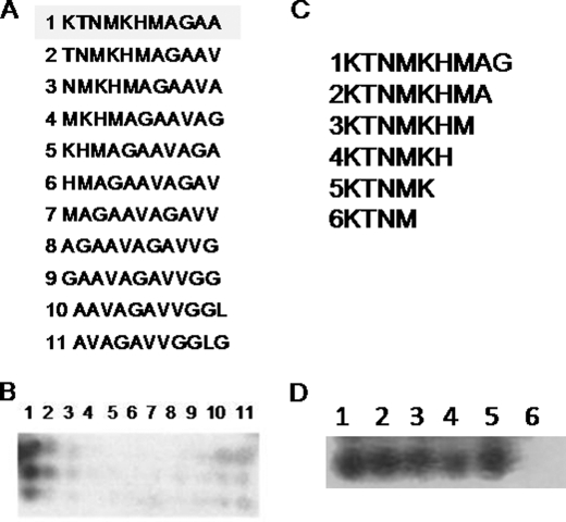
FIGURE 3.
Mapping PrP-AA binding epitopes. Domain specificities of PrP-AA were determined using a peptide microarray. Sequences of either sequentially one amino acids shifted (A) or single amino acids deletions (C) peptides within region PrP106–126 which were synthesized and spotted on membranes are displayed in A and C. Membranes were then probed with PrP-AA (2 μg/ml) and then HRP conjugated anti-human-IgG antibody (triplicate membranes were probed). The sequence motif KTNMK appeared to be highly important since only peptide 1 is bound by PrP-AA, as shown in panel B. Further validation came from experiments shown in panel D, which show strong binding only when residues 1–5 are present, implying the two lysines (KXXXK) are key elements for binding.

