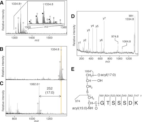
FIGURE 2.
Lyso structure of lipoprotein from B. cereus. MALDI-TOF MS spectra of a fraction containing in-gel-digested PrsA lipoprotein of B. cereus (A) or of the fraction incubated with lipoprotein lipase for 0 (B) or 116 h (C). A deacylated product ion is highlighted. The inset in A is the magnified view. The open circle and closed square indicate the monoisotopic lipopeptide ion containing two fatty acids with total number of double bonds of 0 and 1, respectively. The cross indicates a sodium-added form of the monoisotopic lipopeptide ion containing two saturated fatty acids. Shown at the right are MS/MS spectrum of the N-terminal lipopeptide ion at m/z 1334.8 (D) and elucidated lyso structure of B. cereus PrsA (E). The y-series ions and product ions that have lost a 17:0 fatty acid or monoacyl(17:0)-thioglycerol are highlighted.

