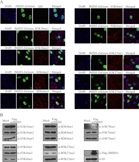
FIGURE 2.
Overexpressed JMJD5 did not affect methylation levels on tested histone H3 lysine residues. A, HEK293T cells were transfected with JMJD5-ZsGreen. After 36 h of transfection, cells were fixed and analyzed by immunocytochemistry. JMJD5 was visualized by the tagged ZsGreen protein (green). Histone H3 lysine methylation levels were observed by staining with the indicated primary antibodies (red). IgG antibody was used for the negative control. DAPI was used to stain nuclei (blue). B, HEK293T cells were transfected with FLAG-mJMJD5. After 48 h of transfection, chromosomal extracts were prepared and analyzed by immunoblotting with the indicated primary antibodies. Histone H3 was used as loading control for immunoblotting.

