FIGURE 4.
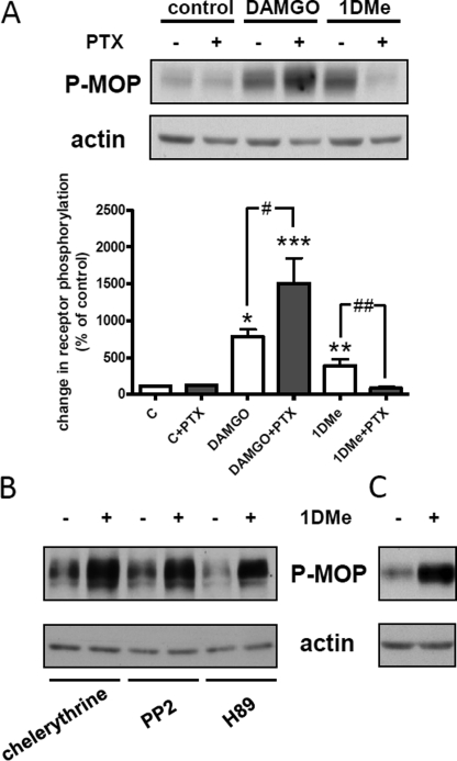
Effect of pertussis toxin and protein kinase inhibitors on MOP receptor Ser-377 phosphorylation. A, representative Western blot showing the levels of Ser-377 phosphorylation induced by treatment with 1 μm DAMGO or 1 μm 1DMe for 30 min in control cells or cells pretreated with 100 ng/ml of PTX for 18 h. Samples were immunoblotted with anti-Ser(P)-377 antibody followed by anti-actin antibody for normalization. Histogram shows the quantification of Ser-377 phosphorylation, and data are expressed as means ± S.E. of three independent experiments. c indicates control cells. *, p < 0.05; **, p < 0.01; ***, p < 0.001 versus untreated cell; #, p < 0.05; ##, p < 0.01; one-way ANOVA followed by Bonferroni post-tests. B, Western blot showing the levels of Ser-377 phosphorylation induced by treatment with 1 μm 1DMe for 30 min in the presence of 10 μm of protein kinase inhibitors (representative of three independent experiments). C, Western blot showing the levels of Ser-377 phosphorylation induced by treatment with 1 μm 1DMe for 30 min in the presence of 10 μm of EGF receptor inhibitor (representative of three independent experiments).

